Keratoconus
Jump straight to:
What is keratoconus?
What are the different types of keratoconus?
What causes keratoconus?
What are the signs and symptoms of keratoconus?
Is keratoconus serious?
How is keratoconus diagnosed?
What are the treatments for keratoconus?
What can help keratoconus?
What research is there into keratoconus?
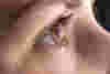
What is keratoconus?
Keratoconus affects the cornea, the clear round-shaped front surface of the eye. In those affected the cornea becomes progressively thinner and weaker over time, eventually causing a cone-shaped protrusion to develop. The change of shape causes blurred and distorted sight as it prevents the light that enters the eye from being correctly focused.
-
- 1 in 500
- people worldwide may have keratoconus - estimates vary between 1 in 500 and 1 in 2,000
What are the different types of keratoconus?
The main types of keratoconus are forme fruste keratoconus (FFK), round cone keratoconus and oval keratoconus. There is also a rare type called posterior keratoconus, and keratoglobus keratoconus, which is a slightly different condition.
-
Forme fruste keratoconus (FFK)
The most common type of keratoconus. It is also the most mild. FFK is usually symptom-free and only diagnosed by mapping the surface of the eye. In FFK the disease starts, but for some reason it stops progressing without any need for treatment.
-
Round cone keratoconus
Round cone keratoconus affects a relatively small area of the cornea, however the steepness of the affected area can be extreme.
-
Oval keratoconus
Oval or sagging cone keratoconus affects a larger portion of the cornea. It can cause rupturing (or hydrops) of the cornea's internal membranes and scarring, as well as making it harder to fit contact lenses.
-
Posterior keratoconus
In posterior keratoconus there is a small region with an increase in curvature at the back of the corneal surface. It's rare and is usually congenital (a condition you are born with) and is unrelated to the other forms of keratoconus.
-
Keratoglobus
Keratoglobus differs from keratoconus. Instead of a single point of thinning of the cornea (which results in a bulging and the development of a cone), the cornea in people with keratoglobus is thin everywhere.
What causes keratoconus?
Scientists do not understand exactly what causes keratoconus. But it is thought that many different genetic and environmental factors are involved. Keratoconus may be more common in certain populations.
Several genes have so far been linked with keratoconus. Although these genetic variants can influence a person’s risk of developing the condition, whether they do so is determined by complex interactions between other genes and environmental triggers.
Scientists are also searching for environmental triggers that may contribute to the development of keratoconus. For example, around one-third of people with the condition also have an allergy. One theory is that allergies can cause itchy eyes – and this may lead to excessive eye rubbing over long periods of time, which could potentially further weaken the cornea.
Keratoconus is also thought to involve a defect in the structure of collagen, the tissue that makes up most of the cornea.
Most people who develop keratoconus have no family history of the condition, and they are otherwise well. But keratoconus can occasionally appear to run in families and also sometimes occur as part of another genetic eye condition, such as Leber congenital amaurosis or retinitis pigmentosa – or a broader inherited syndrome affecting many parts of the body such as Down’s, Marfan or Ehlers-Danlos syndromes.
Dr Nasser Siabi, OBE, is one of five brothers. Of those, four had keratoconus. Listen to the podcast where he discusses his family history and talks about assistive tech.
What are the signs and symptoms of keratoconus?
Your eyesight will often be unaffected in the very early stages of keratoconus, although it may be slightly blurred and distorted. As the condition progresses, your sight may become more distorted due to further bulging of the cornea, which will eventually lead to further short-sightedness and astigmatism.
You will often need frequent changes to your glasses or contact lens prescription to correct this. You may also have increased sensitivity to glare and might see halos around bright lights, which can affect your ability to drive at night. When the condition is more advanced, you may have scarring of the cornea that can blur the vision further.
Keratoconus can affect each eye differently. You may initially only be affected in one eye but eventually, both corneas usually develop the condition – although this may not be with the same severity.
Keratoconus typically begins in young people during their teens or early 20s, with the changes in the eye slowly getting worse over time. Their symptoms will stabilise when the shape of their cornea stops changing, which usually happens around twenty years later.
Is keratoconus serious?
Currently there is no cure for keratoconus, although newer tools like cross linking can prevent further progression if the disease is detected early. There are tools to manage the condition. In the early stage of keratoconus, glasses can be used to give a good standard of vision.
Most people with keratoconus can achieve good sight with the right contact lenses. But in a few severe cases of keratoconus, a person may need a corneal transplant to correct their sight. This involves surgery to remove all or part of a damaged cornea and replace it with healthy tissue from a donor. Even after a corneal transplant, a person almost always still needs to wear spectacles or contact lenses to correct their vision.
How is keratoconus diagnosed?
It’s important to have regular eye tests so that keratoconus is diagnosed as early as possible and for monitoring the progression of the condition. This will help ensure that you receive the right prescription to maintain good sight, as well as access to collagen cross-linking which may help stop your keratoconus from getting worse.
Stay in the loop
on eye research breakthroughs, inspiring real life stories and more...

What are the treatments for keratoconus?
When keratoconus is in its early stages, your eyesight can usually be corrected with spectacles or soft contact lenses. When the condition is more advanced, rigid gas permeable contact lenses are usually needed, and different contact lenses are considered as keratoconus progresses and the cornea becomes more irregular in shape.
Each person's contact lens fit can change over time. Optometrists can help with choosing the best kind and ordering a new type of contact lens if required. Newer treatments have also become available to stop the condition from getting worse.
You can't have laser vision correction if you have keratoconus.
The main treatments for keratoconus are:
Corneal cross linking
Corneal cross linking is a newer treatment which is an option if keratoconus is detected at an early stage when it's still progressing. It involves removing a small area of the surface of the cornea, and applying drops of vitamin B2 (riboflavin) followed by a burst of ultraviolet-A (UV-A) light.
This is thought to cause the tiny collagen fibres in the cornea to join together, making it stronger to stop further deterioration. It does not reverse existing changes.
Custom soft contact lenses
Custom soft contact lenses are intended for mild to moderate keratoconus. Lenses need to be replaced every three to six months depending on the material and your reaction to the lens. Soft lenses are easier to get used to than rigid lenses but might not give such a crisp image.
Rigid gas permeable (RGP) contact lenses
Hard lenses are made of a material which allows oxygen to pass through it. RGP lenses are often a little uncomfortable at first but this becomes less as time goes by - if discomfort continues, other types of contact lenses can be considered.
"Piggybacking" contact lenses
The piggyback system consists of a rigid gas permeable lens sitting on top of a soft contact lens. This combination can lead to clear vision without the discomfort which can come from wearing hard lenses - the soft contacts act as a shield and a cushion to provide additional comfort.
Piggybacks can be expensive, as this can involve two different cleaning systems and soaking solutions.
Hybrid contact lenses
In hybrid contact lenses, the centre is a rigid contact lens (to offer best vision) and the outer part is a soft contact lens, to offer more comfort. These lenses can be difficult to fit and many people find them very difficult to put in and take out.
Scleral and semi-scleral lenses
Scleral lenses are larger diameter lenses that lie on the white part of the eye (the sclera) and float above the cornea without touching its surface. People who have advanced keratoconus or extremely protruding cones could benefit from this option.
In general, these lenses are very comfortable to wear and provide excellent vision in select patients who cannot tolerate other types of lenses.
Corneal ring implants
Corneal ring implants (such as Intacs) are thin plastic, semi-circular rings that are inserted into the mid layer of the cornea. A local anaesthetic is used to perform the procedure, which helps to reshape the cornea by flattening it to improve vision.
This can improve uncorrected vision in most patients, although glasses or contact lenses may still be needed for functional vision.
People who should not have corneal ring implants include pregnant women, people who have other eye health problems that may cause future problems, or people taking certain medication that may impair healing of the eye.
Topography-guided conductive keratoplasty
Topography-guided conductive keratoplasty is a non-invasive treatment that uses radiofrequency to send radio waves to specific areas on the cornea. This is administered by a handheld probe and could be effective in reshaping corneal configuration in people with keratoconus.
Corneal transplant
Most people with keratoconus can achieve good sight wearing contact lenses. But in a few severe cases of keratoconus, a person may need a corneal transplant to correct their sight.
Often referred to as keratoplasty or a corneal graft, a corneal transplant is an operation to remove all or part of a damaged cornea and replace it with healthy donor tissue. Keratoconus is one of the most common reasons for a cornea transplant.
The procedure can be carried out under general anaesthetic or local anaesthetic. It usually takes less than an hour and you'll either leave the hospital the same day or stay overnight. It can take 12-18 months for the vision to fully recover and further surgery may be required to achieve the best outcome. Even after a corneal transplant, a person almost always still needs to wear spectacles or contact lenses to correct their vision.
The type of corneal transplant you have will depend on which part of the cornea is damaged or how much of the cornea needs replacing. The options include penetrating keratoplasty (PK), which is a full-thickness transplant, and deep anterior lamellar keratoplasty (DALK) which is replacing or reshaping the outer and middle (front) layers of the cornea.
What research is there into keratoconus?
Studying the function of genes which are known to be involved is helping scientists to improve our understanding of the biology of keratoconus – and will hopefully lead to new prevention or treatment strategies. They have so far identified that these genes are involved in many diverse activities – including in eye development, the formation and structure of the cornea, the lattice of materials that fill spaces between the cells of the cornea, inflammation and in the control of cell growth.
One of Fight for Sight’s strategic programmes invests in research into eye diseases like keratoconus.
Fight for Sight is currently funding Dr Veronique Vitart and her team at the University of Edinburgh who are investigating corneal thinning to ultimately reduce increasing demand on corneal transplants. The focus of the study will be on keratoconus as well as Brittle Cornea Syndrome (BCS), a rare but devastating genetic disorder.
Previously, we funded Dr Mouhamed Al-Aqaba and his team of researchers at Nottingham University who were using biological ‘markers’ to investigate the underlying nerve structures in keratoconus.
Fight for Sight also previously contributed to research at Guy's Hospital and Moorfields Eye Hospital to understand more about the effectiveness of corneal cross-linking.
Our research is fuelling projects helping to unlock the secrets of dozens of different eye conditions. The brilliant minds we fund are working to understand how eye conditions start, how to prevent them, and to diagnose them sooner. And they’re finding new treatments.
With your help we’ll use our expertise to prevent, treat and cure vision loss within a lifetime.

Last updated November 2022
Approved by Prof Stephen Tuft, Moorfields Eye Hospital NHS Foundation Trust
-
Understanding
-
Diagnosis
-
Treatment
-
Our research
- Understanding
- Diagnosis
- Treatment
- Our research